Part XIII: Albert Claude, 1974 Prize in Physiology or Medicine.
By Joseph Luna
On December 7, 1970, the moon-bound crew of the final Apollo mission swiveled their camera toward earth, some 28,000 miles distant, and took a picture. Three weeks later the resulting photograph revealed a delicate blue orb suspended in space, painted with swirling clouds above the African continent. When released to the public in time for the holiday newspapers, this picture became instantly famous, serving as a visual capstone for humanity’s sojourn beyond our planet, which appears simultaneously majestic and intimate. It is perhaps for that reason that this picture was dubbed “The Blue Marble,” and is among the most iconic scientific photographs known.
I wonder what our next three prize-winners thought of the Blue Marble photo that winter. Whereas astronauts helped make the world small with spectacular portraits of earth, by the 1970s our next three Scandinavian visitors, Albert Claude, George Palade, and Christian de Duve, had been using images for over 25 years to show that microscopic cells were organized worlds unto themselves. Starting with the first electron microscope image of an intact cell in 1945, these three (and many others) helped launch the modern discipline of cell biology. For a comprehensive history of cell biology, particularly at Rockefeller University, I refer the reader to “Entering an Unseen World” by our very own Carol Moberg. For the next three installments of this series, we’ll specifically profile how each of these three men contributed to found a field as a distinct RU creation. And we’ll begin with Albert Claude.
Claude’s early life was difficult, and a bit momentous. After losing his mother to breast cancer at the age of seven, Claude moved around with his family before dropping out of school to care for an ailing uncle. He never finished high school. He worked in a steel mill during World War I, and volunteered as a teenager to aid the British Intelligence Service. By the war’s end, Claude was a decorated military veteran, and his first lucky break came when Belgian education authorities made it possible for veterans to pursue higher education without a diploma. This made it possible for Claude to go to medical school in 1922 and he graduated six years later.
It was then that Claude turned his attention to the cancer problem. At the time, The Rockefeller Institute for Medical Research (RIMR) was an epicenter for the debate on the origin of cancer. On one side was Peyton Rous, discoverer of the first transmissible sarcoma in chickens that bears his name, as the chief proponent for a viral origin of cancer. On the other side was James Murphy, who in short believed that a chemical or environmental insult was responsible for inducing cancer in otherwise normal cells. What exactly the Rous sarcoma agent was could only be speculated, since few had tried to purify it. Claude, freshly read up on the subject, wrote to then RIMR president Simon Flexner and proposed isolating the sarcoma agent. A year later Claude found himself in Murphy’s laboratory in New York, charged to do just that.
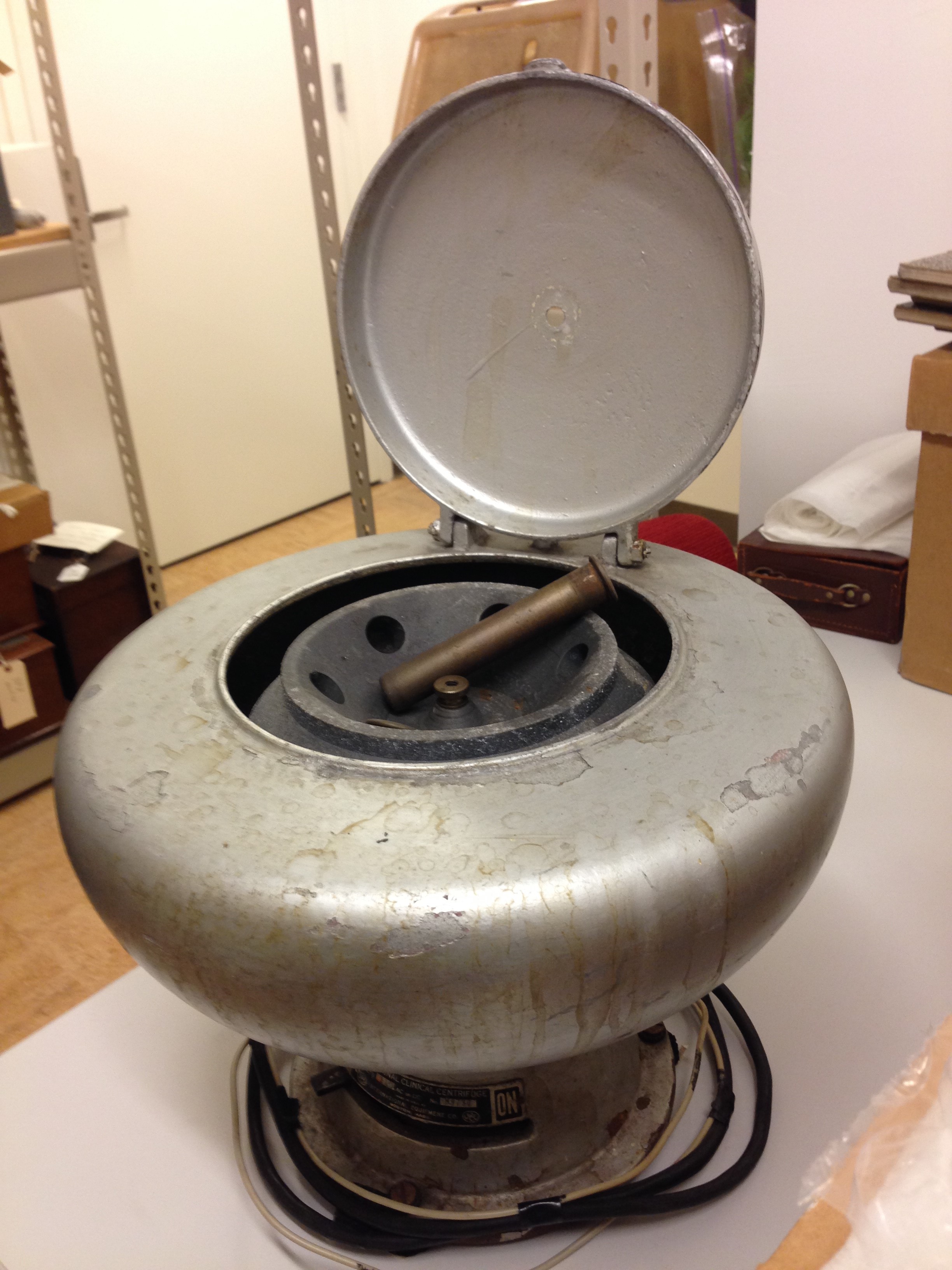
Claude initially attempted to purify the tumor agent via chemical means, but met numerous problems. The basic idea for each experiment was to test if the tumor agent could be concentrated, which, if so, meant that it should be more potent at inducing tumors. But chemical means didn’t work consistently. Taking a cue from researchers in England, Claude opted to try and physically separate the tumor agent using newly available centrifuges. By taking ground up cancer cells and placing them in a centrifuge, Claude was able to physically separate cell contents on the basis of differing size and mass. He could then carefully test each layer of liquid (called a fraction) in the centrifuged tube to see if it could still induce tumors. By tinkering with the speed and order of spins with a centrifuge, Claude could thus physically separate known subcellular structures into four rough fractions: large and heavy nuclei, medium-sized mitochondria, a smaller fraction he eventually termed “microsomes,” and the freely soluble cytoplasm. Moreover, he could subject each fraction to a battery of chemical, biological and microscopic tests. This achievement, to fractionate cells on the basis of differential centrifugation, became the first essential technique for cell biologists, since it allowed one to test where a particular activity (like that of an enzyme or a virus) was roughly located in the cell. And as a control, Claude also fractionated normal, non-cancerous cells, and achieved similar results. Since these normal cells had no tumor agent they couldn’t cause cancer. On the basis of how similar they fractionated compared to cancer cells, Claude and Murphy reasoned that what they first needed was a definition of a “normal” cell. What was initially a biomedical question on cancer, had turned into something much more basic: what did normal cells look like and how did they work?
With his fractionation technique, Claude broke open cells and did his tests, which often included observing the contents of each fraction in the microscope. It stood to reason that for smaller and smaller things, from mitochondria to microsomes, he would need a more powerful microscope. Upon publishing an article in 1943 on what he observed in cytoplasm using his fractionation methods, Claude received a letter from the Interchemical Corporation in New York, proposing the use of their new electron microscope to observe Claude’s cell fractions. The rest as they say, is history: the unusual collaboration between academic scientists and a commercial research laboratory yielded extraordinary dividends that culminated in cell biology’s own Blue Marble: the first EM picture of a whole cell in 1945, taken by Albert Claude, Keith Porter and Ernest Fullam.
As much as we’d like to think otherwise, the primary purpose of the last Apollo mission was to do the painstaking work of furthering lunar geology and testing the limits of human hardware in space; the Blue Marble was a stirring but side outcome. Just the same, even though we credit Albert Claude in part for taking the first EM picture of a cell, no less stirring, it was the meticulous fractionation methods he pioneered that formed his key contribution to new and exciting views of the world inside cells.